Case Report - (2022) Volume 9, Issue 1
A conventional technique for a migrated JJ Stent in an infant
Jayalaxmi Shripati Aihole*Abstract
Indwelling ureteral stents have been used temporarily in a variety of urological procedures for many years in adults, children as well as in infants. However their use is not without complications, therefore they should be ideally removed once their intended purpose is served in 6 to 8 weeks. One such untoward scenario, the proximal migration of JJ stent is though not uncommon, is challenge to retrieve. Author is describing here a conventional retrograde cystoscopic technique for its retrieval.
Keywords
Migrated double J stent, proximal migration of ureteric stent, guide wire, cystoscope, JJ removal forceps
Highlights
What is known: Use of JJ Stent is not without complications; therefore they should be ideally removed once their intended purpose is served in 6 to 8 weeks.
Among these methods, ureteroscopy with the use of grasping forceps, helical basket and ureteral balloon dilator tip have been described in adults and last option may be antegrade approach via nephroscopy including nephrostomy.
What is unknown: Choosing the adequate length and calibre with proper lubrication and gentle and easy passage of a JJ stent over a guide wire intra operatively, and confirmation of both tips in respective position by imaging can possibly avoid such migration.
The retrieval of proximally migrated JJ stents in infants is technically challenging procedure. However, with adequate available instrumental facility and imaging guidance, a conventional retrograde technique can be implanted without morbidity in a tertiary care paediatric and neonatal centre.
Introduction
JJ stents are commonly used for variety of urological conditions for relieving the obstruction, diversion, and for reconstructive procedures in adults as well as in children. Though their use in neonates has rarely been documented in literature especially in neonatal pyeloplasty. Once their purpose is served they should be removed, since they are associated with complications. Conventionally retrograde cystoscopic route is the preferred route for removal of stents, however, may not be feasible in proximal migration. Antegrade percutaneous nephrostomy and removal of the ureter stent can be used such cases and is well described in the literature.
Case Report
A 45 days old male neonate born by full term vaginal delivery with a birth weight of 2.8 kg, to a non consanguineously married couple was referred to us with a history antenatally detected at 28 weeks left hydronephrosis. Baby had right congenital talipus equinovarus deformity for which was serial plaster cast was applied.
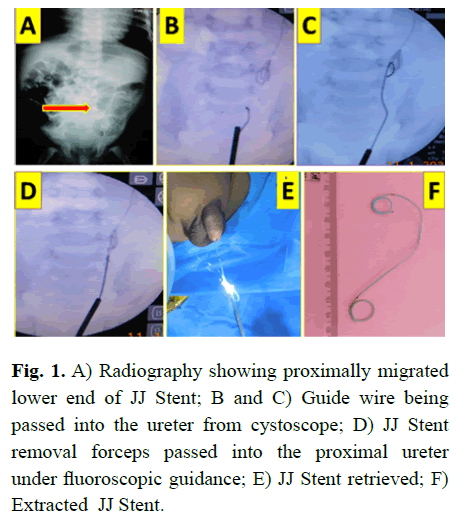
Bay was evaluated by technetium 99 m Ethyelenedicysteine (EC) scan which proved left pelvi uretric junction obstruction with 19% differential function. Hence left modified Anderson Hynes dismembered pyeloplasty with insertion of 2.8 fr JJ Stent across. Once the baby came for planned JJ stent removal after 6 weeks, radiography showed proximally migrated JJ in the almost at pelvi-uretric junction and upper ureter (Fig 1).

Figure 1: A) Radiography showing proximally migrated lower end of JJ Stent; B and C) Guide wire being passed into the ureter from cystoscope; D) JJ Stent removal forceps passed into the proximal ureter under fluoroscopic guidance; E) JJ Stent retrieved; F) Extracted JJ Stent.
Under general anaesthesia in lithotomy position, 9.5 fr cystoscope passed into bladder, left ureteric orifice was cannulated with 0.38/0.97 mm guide wire comfortably and replaced with JJ Stent removal forceps; and JJ stent was grasped with its jaws and under fluoroscopic guidance throughout, JJ was removed absolutely uneventfully within few minutes and baby was discharged after 6 hours (Fig 1).
Results and Discussion
JJ Stents are soft hollow flexible tubes having curls at both ends, comes in various sizes and lengths, used temporarily in urological practise since years to relieve the obstruction, diversion and for reconstructive procedures in adults as well as in children to allow free flow of urine across the ureter. Short term sequelae may include pain, hematuria, dysurea, and stent migration. Whereas, the long-term sequelae from ‘‘forgotten’’ stents include occlusion, encrustation, fragmentation, extrusion, abscess formation, renal failure, and sepsis which carry even greater morbidity. Therefore JJ stent generally needs to be replaced or removed in 6 to 8 weeks. The incidence of ureteric stents migrating proximally is reported as 2% [1-3].
The use of JJ Stents in infants and neonates has rarely been described especially in the literature.
Though various methods of retrieval for such migrated stent have been described in adults, however, are technically challenging in children as well as in infants due to the structural anatomy [4-6].
Among these methods, ureteroscopy with the use of grasping forceps, helical basket and ureteral balloon dilator tip have been described in adults and last option may be antegrade approach via nephroscopy including nephrostomy[3,5].
Ours is a tertiary care paediatric and neonatal care institute in southern India where we conduct neonatal and infantile pyeloplasties with smaller calibre JJ stents.
One such rare incident has been reported here, wherein during neonatal left pyleoplasty 2.8 Fr JJ stent was placed intra operatively and baby was kept one regular follow up. Imaging studies on follow up confirmed gradual proximal migration JJ stent without any symptoms (Fig 1).
Hence after 6 weeks, under general anaesthesia in lithotomy position under fluoroscopic guidance proximally migrated JJ Stent was extracted with routine 9.5 Fr cystoscope and JJ removal forceps with ease uneventfully.
Conclusion
Choosing the adequate length and calibre with proper lubrication and gentle and easy passage of a JJ stent over an appropriate guide wire intra operatively, and confirmation of both tips of JJ in respective position in renal pelvis and bladder by imaging can possibly avoid such migration. The retrieval of proximally migrated JJ stents in infants is technically challenging procedure.
However, with adequate available instrumental facility and imaging guidance, a conventional retrograde technique can be attempted without morbidity in a tertiary care paediatric and neonatal centre.
Conflicts of Interest
Disclosure of potential conflicts of interest-NONE
Disclosure
Research involving Human Participants and/or Animals-NO
Financial Disclosure
Funding-nil
Acknowledgement
Author would like to thank all her pediatric surgeical collegues, anesthetists, ot staffs, and radiology staffs of igich bengaluru, karnataka.
Manuscript has not been previously published in whole or in part or submitted elsewhere for review.
Consent
Informed consent-verbal as well as written consent taken from parents.
References
- Aihole JS, Muniyappa NB, Javaregouda D, et al. Forgotten JJ stent: A rare case report. Ped Urol Case Rep. 2015; 2:6-10.
[CrossRef], [Google Scholar]
- Aihole JS, Babu MN, Jadhav V, et al. Neonatal giant hydronephrosis: A rare case report. Afr J Uro. 2018; 24:126-9.
[CrossRef], [Google Scholar]
- Pérez-Bertólez S, Alonso V. Removal of an intra-renal migrated ureteral stent through a percutaneous nephroscopy in a 2-year-old child. Urol Case Rep. 2021; 34:101482.
[CrossRef], [Google Scholar] [Pubmed]
- Jayakumar S, Marjan M, Wong K, et al. Retrieval of proximally migrated double J ureteric stents in children using goose neck snare. J Indian Assoc Pediatr Surg. 2012; 17:6.
[CrossRef], [Google Scholar] [Pubmed]
- Shin JH, Yoon HK, Ko GY, et al. Percutaneous antegrade removal of double J ureteral stents via a 9-F nephrostomy route. J Vasc Interv Radiol. 2007; 18:1156-61.
[CrossRef], [Google Scholar] [Pubmed]
- Boardman P, Cowan NC. Technical report: Fluoroscopically guided retrograde ureteric stent retrieval and replacement using a guide catheter directed snare. Clin Radiol. 1997; 52:308-9.
[CrossRef], [Google Scholar]
Author Info
Jayalaxmi Shripati Aihole*Received: 02-Feb-2022, Manuscript No. PUCR-22-52860; , Pre QC No. PUCR-22-52860(PQ); Editor assigned: 04-Feb-2022, Pre QC No. PUCR-22-52860(PQ); Reviewed: 16-Feb-2022, QC No. PUCR-22-52860; Revised: 21-Feb-2022, Manuscript No. PUCR-22-52860 (R); Published: 28-Feb-2022, DOI: 10.14534/j-pucr.2022.288
Copyright: This is an open access article distributed under the terms of the Creative Commons Attribution License, which permits unrestricted use, distribution, and reproduction in any medium, provided the original work is properly cited.
