Case Report - (2022) Volume 9, Issue 2
A Huge abdominal cystic mass: Rare cause for crossed renal ectopia
Thayer M. Aboush*, Zaidoon Moayad Altaee and Mahmood Mosa MahmoodAbstract
Crossed renal ectopia is a rare congenital anomaly of the renal system in which most cases are asymptomatic and discovered incidentally. In case presentation we report an unusual case of a 2.5-year-old-male child a known case of cerebral palsy who diagnosed previously with agenesis of left kidney he presents with gradually developed abdominal mass The clinical suspicion and MRI was that of a mesenteric cyst or cystic teratoma but during operation an ectopic non fused left kidney discovered and nephrectomy of the left non-functioning kidney done Conclusion. Crossed ectopic kidney although it is uncommon cause of abdominal mass it should be suspected in patient with abdominal cystic mass especially in case of absent kidney in the other side.
Keywords
Crossed renal ectopia, abdominal cystic mass, kidney
Introduction
Crossed renal ectopia is one of the rare congenital anomalies of the urinary system in which the ectopic kidney crosses the mid line to be located contralateral to its normal position while its ureter inserted to the bladder in its normal side [1,2]. Most cases remain asymptomatic, and only 20%-30% are incidentally discovered during autopsy, screening tests, or by investigations for other abdominal pathologies [1,2]. Crossed renal ectopia has male predominance of 3:2 to 2:1 [2,3]. Right to left ectopia is two to three times more frequent than left to right variety [4-6].
The kidneys are fused in about 85%-90% of cases and its incidence is about 1:7500 autopsies, while non-fused variety has incidence of 1:75000 autopsies and represent 10% of cases [2,3,7] .
Case Presentation
A 2.5-year-old-male child a known case of cerebral palsy presented with right side abdominal distention which developed gradually over more than six months, also he had anorexia, constipation, weight loss and vomiting sometimes, with recurrent attacks of urinary tracts infections.
On physical examination an eight kg male with normal vital signs has asymmetrical abdominal distention mainly in the right side and on palpation a mass detected extending from the right subcostal margin down to the pelvis and medially it was extended to the midline it was soft in nature, fixed, not tender and dull on percussion (Fig 1).
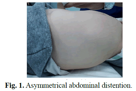
Fig 1: Asymmetrical abdominal distention.
Complete blood count was normal a part from lymphocytosis, renal function test was normal, U/S revealed a huge cystic mass involving the right abdominal cavity with right sever hydronephrosis and the left kidney was not visualized, I.V.U show right kidney hydronephrosis with kinking of ureter and no excretion in the left kidney (Fig 2).
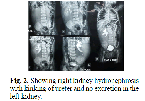
Fig 2: Showing right kidney hydronephrosis with kinking of ureter and no excretion in the left kidney.
M.R.I of abdomen was done and the report was that: the right kidney has mild hydronephrosis and the left kidney not seen (agenesis) and there is a large thin wall cystic mass occupies most of the right abdomen (11.5 cm* 19.5 cm*10 cm) looks retroperitoneal causing distortion of the right kidney suggest benign lesion, cystic teratoma or mesenteric cyst (Fig 3).
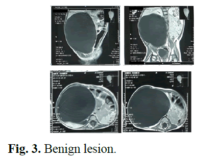
Fig 3: Benign lesion.
During exploration the left kidney identified Fig 4, is to be in the right side just overlay the right kidney and it was huge in size sac-like making pressure effect on the right kidney, and it was malrotated, so the pelvis was anteriorly directed and the ureter descend down crossing the midline to be inserted in its normal side (Fig 5). There was a clear line of separation in between the two kidneys (Fig 6), decision to do nephrectomy was made because, it looks nonfunctioning kidney from the preoperative investigations and the gross appearance of sac like structure. Nephrectomy done and the patient did very well postoperative, the histopathology result reveals that its case of uretero-pelvic junction obstruction, all investigations was normal post-operative and the pressure effect resolve (Figs 4-6).
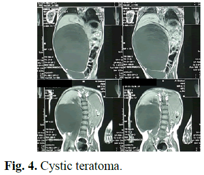
Fig 4: Cystic teratoma.
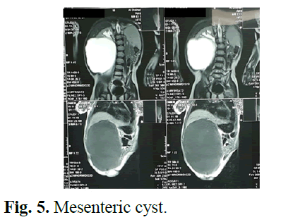
Fig 5: Mesenteric cyst.
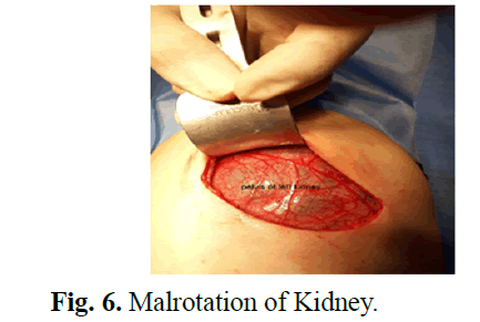
Fig 6: Malrotation of Kidney.
Results and Discussion
Crossed renal ectopia is thought to be occur as a result of abnormal migration and traveling of the metanephric blastoma and ureteric bud cross the midline during the 4-8 weeks of fetal development, or other causes like due to abnormal position of the umbilical artery which thought to be a cause by preventing normal cephalic migration, Malrotation of the caudal end of the fetus, genetic and teratogenic factors are also blamed [6].
Renal ectopia is either simple renal ectopia or crossed renal ectopia and it represent the second most common anomaly after horse-shoe kidney [4].
There are four different variants of crossed renal ectopia: Crossed fused renal ectopia (represents the majority of cases 90%), Unfused ectopia (is un common), Solitary crossed renal ectopia (are very rare) and Unfused bilateral crossed renal ectopia (are also very rare). [1,4,8], and there are six subtypes of crossed fused renal ectopia: inferior, sigmoid or S-shaped, lump, disc, L-shaped and superior crossed fused renal ectopia [4,6].
Understanding the classification and anatomy of each variant of crossed renal ectopia is important in management of symptomatic cases [6].
Crossed renal ectopia usually asymptomatic and discovered incidentally [1,2], and when it is symptomatic the pain or palpable mass will be the most common presenting symptom and also it is may present as urinary tract infection, nephrolithiasis, hydronephrosis, uretero-pelvic junction obstruction as in our case which present’s too late and there was a little renal parenchyma in a non-functioning sac like kidney, Other presentations may be gastrointestinal symptoms, hypertension and renal failure [6,9].
Crossed renal ectopia can be associated with other genitourinary abnormalities like hypospadias, megaureter, cryptorchidism, urethral valve, cystic dysplasia and unilateral agenesis of ovaries and fallopian tube, and non-renal anomalies adrenal, cardiac, gastrointestinal, and skeletal anomalies, it may be associated with genetic syndromes such as (turner syndrome, Edward syndrome, and trisomy-9) [1,5,9,10].
Ultrasound is the initial investigation in the diagnosis of crossed renal ectopia, it could identify an empty renal fossa and the position of the ectopic kidney in the pelvis, subdiaphragmatic area, or even in contralateral side, IVP is usually needed to make the diagnosis, Other diagnostic tools: Are antegrad or retrograde pyelography, nephrotomography, Computerized Tomography (CT) scan, gadolinium-enhanced MR urography, technetium-99 mercaptoacetyltriglycin-3 and arteriography in selected cases [2,5,9,10].
Conclusion
There is no special treatment for crossed renal ectopia when it is asymptomatic and the treatment is indicated when there is complications and when symptoms are present, and the management is according to the presence of complication after thorough clinical and radiological assessment and understanding the anatomy and the cause of pathology. Crossed renal ectopia needs treatment if it’s symptomatic and should be suspected in patient who has been diagnosed as a case with renal agenesis when he complains of other symptoms.
Conflict of Interest
The authors declare that we have no competing interests.
References
- Ratola A, Almiro MM, Lacerda Vidal R, et al. Crossed renal ectopia without fusion: an uncommon cause of abdominal mass. Case Rep Nephrol. 2015; 2015: 679342.
[CrossRef] [Google Scholar] [PubMed]
- Ramaema DP, Moloantoa W, Parag Y. Crossed renal ectopia without fusion-an unusual cause of acute abdominal pain: a case report. Case Rep Urol. 2012; 2012: 728531.
[CrossRef] [Google Scholar] [PubMed]
- Kodama K, Takase Y, Tatsu H. Management of ureterolithiasis in a patient with crossed unfused renal ectopia. Case Rep Urol. 2016;2016: 1847213.
- Degheili JA, AbuSamra MM, El-Merhi F, et al. Crossed unfused ectopic pelvic kidneys: a case illustration. Case Rep Urology. 2018; 2018: 7436097.
[CrossRef] [Google Scholar] [PubMed]
- Al-Hamar NE, Khan K. Crossed nonfused renal ectopia with variant blood vessels: a rare congenital renal anomaly. Radiol Case Rep. 2017; 12:59-64.
- Sadiqi J, Rasouly N. Cross fused renal ectopia with uretero-pelvic junction obstruction in upper moiety and renal pelvis stones in the lower moiety. Mathews J Case Rep. 2016;1:1-3.
- NeMoyer R, Narayanan S, Maloney-Patel N. A case of crossed left renal ectopia identified during colostomy reversal. Case Rep Surg. 2017; 2017: 3674603.
[CrossRef] [Google Scholar] [PubMed]
- Al Mugeiren MM. Association of multicystic dysplasia and crossed nonfused renal ectopia: a case report. Saudi J Kidney Dis Transpl. 1997; 8(2):148.
[Google Scholar] [PubMed]
- Felzenberg J, Nasrallah PF. Crossed renal ectopia without fusion associated with hydronephrosis in an infant. Urology. 1991; 38(5):450-2.
[CrossRef] [Google Scholar] [PubMed]
- Nursal GN, Büyükderelİ G. Unfused renal ectopia: a rare form of congenital renal anomaly. Annals Nuclear Med. 2005;19(6):507-10.
Author Info
Thayer M. Aboush*, Zaidoon Moayad Altaee and Mahmood Mosa MahmoodReceived: 31-Mar-2022, Manuscript No. PUCR -22- 59219; , Pre QC No. PUCR -22-59219 (PQ); Editor assigned: 06-Apr-2022, Pre QC No. PUCR -22-59219 (PQ); Reviewed: 27-Apr-2022, QC No. PUCR -22-59219; Revised: 06-May-2022, Manuscript No. PUCR -22-59219 (R); Published: 16-May-2022, DOI: 10.14534/j-pucr.20222675577
Copyright: This is an open access article distributed under the terms of the Creative Commons Attribution License, which permits unrestricted use, distribution, and reproduction in any medium, provided the original work is properly cited.
