Case Report - (2021) Volume 8, Issue 3
Paediatric Posterior Urethral Polyp: A Case Report
Rahul. K. Gupta, Manishkumar P. Khobragade*, Beejal.V. Sanghvi, Kedar.P. Mudkhedkar, Deepa.P. Makhija, Dr. Rujuta.S. Shah, Soundharya.S and Sandesh.V. ParelkarAbstract
Urethral polyps also known as fibroepithelial polyps, prostatic urethral polyps (in males) or benign urethral polyps and are very rare. They are most common benign mesodermal tumours of the urinary tract. They present with variety of urinary tract symptoms. Voiding Cystourethrogram (VCUG) is the first investigation requiring careful interpretation. Cysto-urethroscopy is the investigation of choice for final diagnosis and excision of urethral polyps. We report a case of an 8-year-old boy who presented with lower urinary tract symptoms. Radiological investigation revealed bilateral hydroureteronephrosis. We confirmed the diagnosis and excised the urethral polyp endoscopically with Holmium YAG Laser rendering patient completely free of symptoms.
References
Blog - Find Lawyer in Utah Real Estate in Philippines Blog | Best Betting Sites in Australia Real Estate in UK Blog | Casino Sites in Ethiopia Real Estate in USA Blog - Find Lawyer in Texas Real Estate in Croiatia Real Estate in Balkans Real Estate in Brazil Blog | Best Betting Sites in China Real Estate in Australia Real Estate in Spain Blog - Find Lawyer in Illinois Blog - Find Lawyer in Colorado Real Estate in France Blog | Info Alt Coins Blog | Reverse Mortgage in USA Real Estate in Japan Real Estate in MexicoKeywords
Urethral polyp, posterior urethra, benign lesion; cystourethroscopy, holmium yag laser
Introduction
Urethral polyp in paediatric population is a rare entity. They are known by variety of names i.e., fibroepithelial polyps, benign urethral polyps and prostatic urethral polyps. They are generally solitary, congenital in nature, common in young boys and arise from posterior urethra. Average age of presentation is 5.2 years [1]. They arise from verumontanum in males and mid urethra in females. Urethral polyps can be incidental findings during cystoscopy or can present with complains of hesitancy, thin urinary stream, urinary retention, haematuria, sudden painful interruption of urinary stream, dysuria, recurrent UTI, Vesicoureteric Reflux (VUR) or enuresis.
Case History
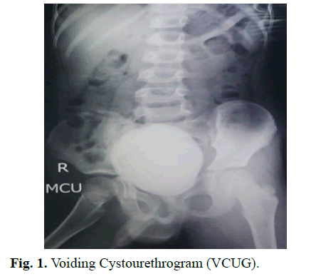
We present a case of an 8-year-old boy with urinary frequency and nocturnal enuresis since 3 years. Urine and blood investigations were normal, sonogram was suggestive of bilateral mild hydroureteronephrosis with cystitis. Voiding Cysto-urethrogram showed mildly dilated posterior urethra with irregular bladder wall with no VUR (Fig 1).

Fig. 1. Voiding Cystourethrogram (VCUG).
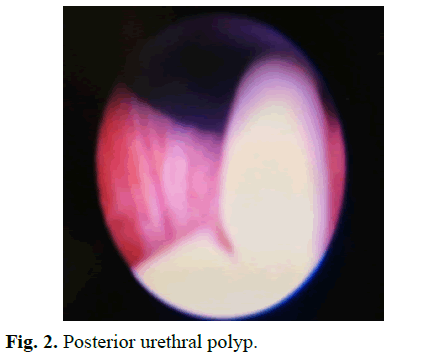
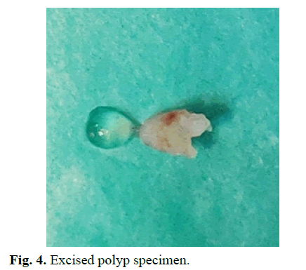
Cystourethroscopy revealed a small 1.5 cm pedunculated polyp arising from the verumontanum going towards the bladder neck. Transurethral resection of the polyp was done with Holmium YAG LASER with energy of 10 Joules, frequency 1 Hz and power of 10 Watts. The specimen was retrieved by DJ holding forceps. Patient showed significant improvement of symptoms from 2nd post-operative day and was discharged on 3rd post- operative day. He is on regular follow up without any symptoms. Histopathology report of specimen showed it to be benign polyp containing fibrous stroma and lined by urothelium.
Results and Discussion
Urethral polyps are rare with only few cases reported so far, from neonate to 70 years of age. Though its incidence is increasing due to better diagnostic techniques and availability of smaller cystoscope exact incidence is not known. Aetiology is controversial; they can be congenital, infective, traumatic and obstructive. Stephens [2] proposed that congenital type may arise as a result of developmental error in the invagination process of the glandular material of the inner zone of prostate. Down [3] considers it as a protrusion of the wall of posterior urethra. Kuppuswami and Moors [4] suggested that it may be due to metaplastic epithelial changes secondary to the oestrogen released during gestation. Inflammatory polyps are considered as a result of UTI however they are uncommon in children [1].
Posterior urethral polyps need to be differentiated from posterior urethral valve, blood clot, polypoid/papillary cystitis, urothelial papilloma, florid cystitis cystica et glandularis, inverted papilloma and rhabdomyosarcoma of the bladder. [5-8].
In early infancy urethral polyp can cause urethral obstruction and in older boys they may be incidental or cause urinary obstruction, haematuria, retention, UTI, enuresis or rarely azotaemia and lower urinary tract symptoms [6,8,9]. Intermittency of symptom with symptom free interval is highly suggestive of urethral polyp. There are two mechanisms of obstruction to urine outflow; a polyp with a long stalk lies inside the membranous urethra and obstructs the lumen of the urethra only during voiding and voiding must be interrupted to allow polyp to return to its normal position; alternately, a large polyp with a short stalk may completely obstruct the lumen (Fig 2). 25% of the patients with urethral polyps have VUR6 [3,10], some can have associated hypospadias, unilateral hydronephrosis and bladder diverticulum.

Fig. 2. Posterior urethral polyp.
VCUG and USG are the most common initial investigations but are not always diagnostic. VCUG may show filling defect in urethra correlating to site of polyp. CT and MRI may be helpful to rule out other causes. Final diagnosis is done by urethrocystoscopy [6,8,11].
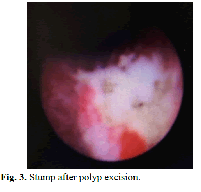
Transurethral resection using LASER or Bugbee electrode is the standard line of management for urethral polyp [8,11]. Larger size polyp can be fragmented into small pieces and can be retrieved from bladder with DJ holding forceps or stone basket (Fig 3 and 4).

Fig. 3. Stump after polyp excision.

Fig. 4. Excised polyp specimen.
Sometimes cystoscopic excision is difficult as they are generally tense, smooth walled and are floating in bladder. Other methods like endoscopic suprapubic approach or open procedures are useful when endoscopic measures fail but urethro-cystoscopy is necessary for confirming diagnosis. The base of the polyp must be fulgurated after excision to prevent recurrence of polyp. K. Fati has reported no recurrence in study of single case of posterior urethral polyps excised by suprapubic cystotomy [6]. Richard Frates has reported recurrence in 1 patient out of 2 cases of posterior urethral polyps excised by suprapubic cystotomy [12]. DeCastro in his study of 17 cases has reported recurrence in one patient excised by cystotomy [10]. Tsuzuki has reported 0% recurrence rate on follow up in 10 cases treated with transurethral resection [5]. Akbarzadeh et al reported transurethral resection in 17 out of 18 patients with no recurrence of follow up of 3 to 17 years [13]. P.Jain has reported a case of cystoscopic multiple posterior urethral polyp excision in an infant with no recurrence [11]. Fatih has reported no recurrence in a patient treated with excision of polyp using Holmium YAG Laser [14].
Histopathologically urethral polyps are benign growths of fibrous connective tissue consisting of smooth muscle core with transitional epithelium covering. Squamous metaplasia and ulceration can be associated [6,8]. Postoperative follow up includes precise history, urinary tract USG and VCUG in patient with VUR.
Conclusion
Though uncommon, urethral polyps should be included in differential diagnosis of any child presenting with symptoms of intermittent lower urinary tract symptoms such as urinary retention or haematuria. Cystourethroscopic excision, preferably with Holmium YAG LASER is the treatment of choice for these polyps with minimum rate of recurrence.
References
- Amogu E, Tamer H, Osama S, Eissa WM, Ghaly MA. Management of male urethral polyps in children: Experience with four cases. Afr J Paediatr Surg. 2009; 6: 49-51.
- Prashant J, Hemanshi S, Sandesh P, Borwankar SS. Posterior urethral polyps and review of literature, Indian J Urol 2007; 23; 206-7.
- Casale AJ. Posterior urethral and other urethral anamoly. Wein AJ, Kavoussi LR, Novick AC, Partin AW, Peters CA, editors. Campbells urology. 49th ed. W.B. Saunders; 2007. 3583-603
- Tsuzuki T, Epstein JI. Fibroepithelial polyp of lower urinary tract in adults. Am J SurgPathol. 2005; 29:460-6
- K. Fathi, A. Azmy, A. Howatson, Carachi R. Congenital posterior urethral polyps in childhood, As Case Report. Eur J Paediatric Surg 2004; 14: 215-7.
- DeCastro R, Campobasso P, Belloli G. Solitary polyp of posterior urethra in children: Report on seventeen cases. Eur J PaedSurg 1993; 3:92-6
- Gary K, Robert L. Alan R. Obstructing polyps of the posterior urethra in boys: Embryology and management. J Urol 1979; 802-804.
- Down RA. Congenital polyp of prostatic urethra: A review of the literature and report of two cases. Br J Urology 1970; 42: 76-85
- Kuppuswami K, Moors DE. Fibrous polyp of the verumontanum. Canad J Surg 1968; 11: 388-390
- Stephens FD. Congenital malformation of rectum, anus and genitourinary tract. Saunders: Philadelphia; 1963; 231.
- Richard F, Frank D. Urethal polyps in male children. 1967; 89: 289-291.
- Fatih O, Ercument K, Turgut Y. Posterior urethral polyp: First holmium-YAG laser ablation on a 3 month-old Infant. J Endourol Case Rep. 2016; 90–92.
- Akbarzadeh A, Khorramirouz R, Kajbafzadeh M. Congenital urethral polyps in children: Report of 18 patients and review of literature. J Pediatr Surg. 2014: 49: 835-839
- Sumit D, William B. Congenital urethral polyps: A rare cause of bladder outlet obstruction in children. Acad J Ped Neonatol. 2017; 2: 555598.
Author Info
Rahul. K. Gupta, Manishkumar P. Khobragade*, Beejal.V. Sanghvi, Kedar.P. Mudkhedkar, Deepa.P. Makhija, Dr. Rujuta.S. Shah, Soundharya.S and Sandesh.V. ParelkarReceived: 19-Apr-2021 Accepted: 03-May-2021 Published: 10-May-2021, DOI: 10.14534/j-pucr.2021267551
Copyright: This is an open access article distributed under the terms of the Creative Commons Attribution License, which permits unrestricted use, distribution, and reproduction in any medium, provided the original work is properly cited.
